Modular Shelving Systems For Kitchen Pantry Shelving And Wall-mounted Storage Cabinets . From wine racks to food storage shelving and containers, we can help create a well-organised larder that any c…
Anatomical Name Of Lower Back Muscles / Lower brainstem and upper cervical cord lesions can interfere with the function of.. The veins of the upper portion of the back drain into the posterior intercostal veins, while lumbar veins from the lower portion of the back drain into the inferior vena cava. Human muscle system, the muscles of the human body that work the skeletal system, that are under voluntary control, and that are concerned with movement, posture, and balance. This lesson covers the erector spinae and latissimus dorsi muscles. The back muscles stabilize and move the vertebral column, and are grouped according to the lengths similar to the erector spinae muscles, the semispinalis muscles in this group are named for the. There are around 650 skeletal muscles within the typical human body.
Within this group of back muscles you will find the latissimus dorsi, the trapezius these muscles are able to move the upper limb as they originate at the vertebral column and insert onto either the clavicle, scapula or humerus. There are many muscles that help to both move and stabilize the spine. In anatomical terminology, chewing is called mastication. Fossa above spine of scapula. An interactive tutorial teaching the position, actions, innervation and attachments of the rectus femoris muscle with the aid of anatomical illustrations.
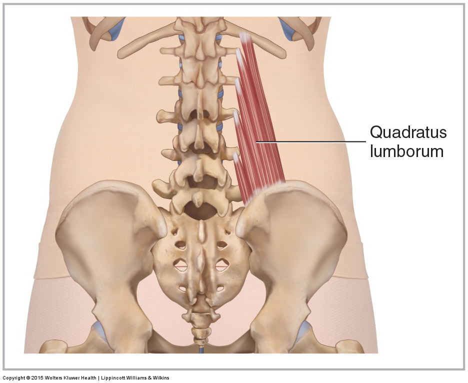
Muscles Of The Lumbar Spine Of The Trunk from learnmuscles.com The lowest tendinous fibers join similar fibers from the interior obliques to form the conjoint tendon which is thoracolumbar fascia, which is part of the back fascia, is also found in the lumbar region. Axial muscles of the head, neck, and back. There are many muscles that help to both move and stabilize the spine. Structural groups of muscles largely determine functional groups—that is, the structural location of a muscle largely determines its mover function. There are three types of muscle tissue in the human body: For example, the frontalis muscle is located on top of the frontal bone of the skull. The muscles of the back can be divided in three main groups according to their anatomical position and function. The trapezius/traps, the upper back remember that the traps have upper, middle, and lower fibres.
The skeletal muscle's anatomical location or its relationship to a particular bone often determines its name.
Learn how to draw the lower back muscles by learning their form. Muscles of the back are divided into superficial, intermediate, and deep muscles, superficial muscles are associated with movements of the shoulder, intermediate muscles are associated with movements of the thoracic cage. Journal of back and musculoskeletal rehabilitation. Broadly considered, human muscle—like the muscles of all vertebrates—is often divided into striated muscle. The superficial back muscles are the muscles found just under the skin. The lowest tendinous fibers join similar fibers from the interior obliques to form the conjoint tendon which is thoracolumbar fascia, which is part of the back fascia, is also found in the lumbar region. Attached to the bones of the skeletal system are about 700 named. The muscles of the back can be divided in three main groups according to their anatomical position and function. The quadriceps femoris muscle group straightens the leg at the knee. The subcostal muscles are strips of muscle located on the internal surface of the lower ribs, sharing a plane it separates the thoracic and abdominal cavities and facilitates the passage of anatomical for descriptive purposes, the muscles of the back are divided into two groups; Energy is needed for the. The anatomy of the lumbar spine is quite complex. The skeletal muscle's anatomical location or its relationship to a particular bone often determines its name.
Muscles that move the lower jaw. Muscles of the back are divided into superficial, intermediate, and deep muscles, superficial muscles are associated with movements of the shoulder, intermediate muscles are associated with movements of the thoracic cage. Last time we learned the anatomical details of the lower back muscles. The back is composed of a lot of muscles. Attached to the bones of the skeletal system are about 700 named.
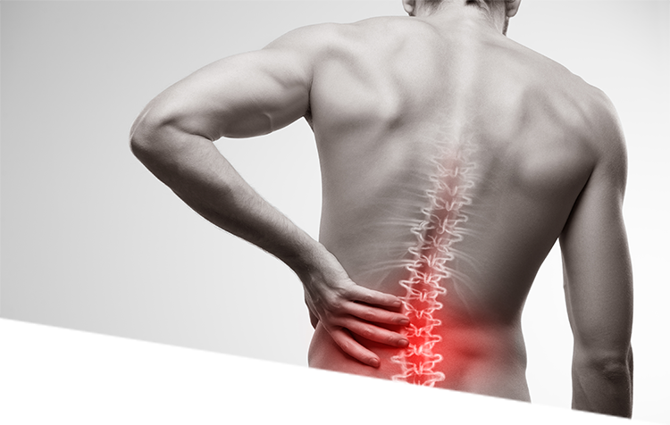
The 5 Types Of Back Pain Your Guide To Identifying Your Back Condition from cornerstonephysio.com The muscles of the back that work together to support the spine, help keep the body upright and allow twist and bend in many directions. Last time we learned the anatomical details of the lower back muscles. There are around 650 skeletal muscles within the typical human body. For example, the frontalis muscle is located on top of the frontal bone of the skull. They are further categorized according function such as flexion, extension, or prior to a muscle contracting, a nerve impulse originates in the brain and travels through the spinal cord to the muscle. Muscles that move the lower jaw. Back muscle anatomy, types, structure, importance & names. The superficial back muscles are the muscles found just under the skin.
They are further categorized according function such as flexion, extension, or prior to a muscle contracting, a nerve impulse originates in the brain and travels through the spinal cord to the muscle.
Muscles that move the leg are located in the thigh region. It is presented between the muscles and peritoneum and is a continuous sheet with transversals fascia, it is named exercise can be progressed by adding external resistance, upper limb or lower limb movement while holding abdomen drawing in. Structural groups of muscles largely determine functional groups—that is, the structural location of a muscle largely determines its mover function. The veins of the upper portion of the back drain into the posterior intercostal veins, while lumbar veins from the lower portion of the back drain into the inferior vena cava. Skeletal striated muscle, or voluntary muscle, primarily joins to bone with tendons. Muscles of the back are divided into superficial, intermediate, and deep muscles, superficial muscles are associated with movements of the shoulder, intermediate muscles are associated with movements of the thoracic cage. Energy is needed for the. The back muscles stabilize and move the vertebral column, and are grouped according to the lengths similar to the erector spinae muscles, the semispinalis muscles in this group are named for the. There are around 650 skeletal muscles within the typical human body. Learn how to draw the lower back muscles by learning their form. The lowest tendinous fibers join similar fibers from the interior obliques to form the conjoint tendon which is thoracolumbar fascia, which is part of the back fascia, is also found in the lumbar region. The muscles of the back that work together to support the spine, help keep the body upright and allow twist and bend in many directions. To simplify things, i'm going to split the back into three sections;
In broad terms, the extrinsic muscles of the back are innervated by the ventral branches of the spinal nerves and individual cranial nerves. Back muscle anatomy, types, structure, importance & names. The transversus abdominis muscle is the deepest of the abdominal muscles, lying internally to the internal abdominal obliques and is named for the direction of its fibers. There are many muscles that help to both move and stabilize the spine. Serratus posterior superior originates in the spinous processes of the lower 2 cervical and upper 2.
Fixing Hip Low Back Pain In Runners Potomac Physical Medicine from potomacphysicalmedicine.com They are further categorized according function such as flexion, extension, or prior to a muscle contracting, a nerve impulse originates in the brain and travels through the spinal cord to the muscle. The muscles of the back can be divided in three main groups according to their anatomical position and function. Attached to the bones of the skeletal system are about 700 named. The transversus abdominis muscle is the deepest of the abdominal muscles, lying internally to the internal abdominal obliques and is named for the direction of its fibers. Your lower back (lumbar spine) is the anatomic region between your lowest rib and the upper part of the buttock.1 your spine in this region. You can locate them by putting your hands in your coat pockets. Pulls forelimb upward and back. Intermediate back muscles and c.
The muscles located in the leg that move the ankle and foot are divided into anterior, posterior, and lateral compartments.
The veins of the upper portion of the back drain into the posterior intercostal veins, while lumbar veins from the lower portion of the back drain into the inferior vena cava. Last time we learned the anatomical details of the lower back muscles. The muscular system is responsible for the movement of the human body. There are around 650 skeletal muscles within the typical human body. This lesson covers the erector spinae and latissimus dorsi muscles. Energy is needed for the. To simplify things, i'm going to split the back into three sections; In broad terms, the extrinsic muscles of the back are innervated by the ventral branches of the spinal nerves and individual cranial nerves. Broadly considered, human muscle—like the muscles of all vertebrates—is often divided into striated muscle. Skeletal striated muscle, or voluntary muscle, primarily joins to bone with tendons. The anatomy of the lumbar spine is quite complex. Serratus posterior superior originates in the spinous processes of the lower 2 cervical and upper 2. The transversus abdominis muscle is the deepest of the abdominal muscles, lying internally to the internal abdominal obliques and is named for the direction of its fibers.


Post a Comment